Anatomy Of Upper Thigh And Hip : Muscles Of The Hips And Thighs Human Anatomy And Physiology Lab Bsb 141 / The upper part of the thigh bone consists of the femoral head, femoral neck, and greater and lesser trochanters.
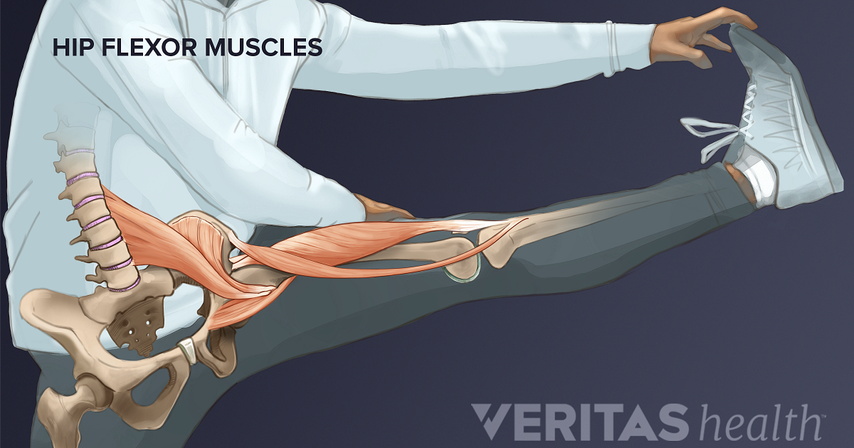
Anatomy Of Upper Thigh And Hip : Muscles Of The Hips And Thighs Human Anatomy And Physiology Lab Bsb 141 / The upper part of the thigh bone consists of the femoral head, femoral neck, and greater and lesser trochanters.. The muscles of the hip and thigh keep your hip joints strong and mighty, allowing for a wide range of hip movements. Iliopsoas muscle, a hip flexor muscle that attaches to the upper thigh bone. Now that you watched the video, you. Hip flexor deep in pelvis a composite o… used to extend the hip when climbing st… Thus, it is thicker in the upper and lateral part of the thigh, where it receives a fibrous expansion from the glutæus maximus, and where the tensor fasciæ latæ is inserted between its layers;
Knee assessment and hip mechanics learn how. Unlike the shoulder girdle, the pelvic girdle is firmly integrated into the axial skeleton: The paired hip bones are connected. Anterior muscles extend your legs and flex your thighs. The information contained in anatomy atlases is not a substitute for the medical care and advice of your physician.
Hip Muscles Anatomy Anatomy Drawing Diagram from i.pinimg.com Most hip fractures sustained by older people result from falls. Mri of upper leg (femur). It is very thin behind and. During hip replacement surgery, your surgeon removes the upper part of your thigh bone, including the femoral head (ball of the hip joint) and a part the upper part of the thigh bone is then exposed, and a series of tools called broaches are introduced one at a time to prepare your thigh bone for a metal. Bones of the lower limb. Want to learn more about it? All of the anatomical parts of the hip work together to enable various movements. The hip's unique anatomy enables it to be both extremely strong and amazingly flexible, so it can bear weight and allow for a wide range of movement.
Muscles of the hips and thighs.
Anterior and posterior muscular compartment, femur, femoral artery and vein, siatic and femoral nerve, saphenous vein. Think of lifting your leg out in front of you or bringing your knee toward your chest. This deep muscle begins in the low back and pelvis and connects on the inside edge of the upper femur. In clinical anatomy the thigh muscles are divided into three groups: It contains many muscles and nerves but only has one bone, the femur, which is the longest and strongest bone in the human body. The hip is a ball and socket joint, formed by the upper thigh bone (the femur) attaching to the acetabulum (the opening pocket in the lower pelvis). During hip replacement surgery, your surgeon removes the upper part of your thigh bone, including the femoral head (ball of the hip joint) and a part the upper part of the thigh bone is then exposed, and a series of tools called broaches are introduced one at a time to prepare your thigh bone for a metal. Muscles of the hips and thighs. It functions to adduct the thigh and to flex and rotate the leg medially at the knee. Anatomy of the thigh : It originates at the anterior inferior iliac spine and just above the acetabulum of the hip bone. Knee assessment and hip mechanics learn how. On the anterior side, the most prominent of the muscles are the sartorius muscle and the four the four muscle of the quadriceps all extend the lower leg, and the rectus femoris additionally can flex the thigh at the hip.
If the iliotibial band (a long tendon that many muscles in your hip and leg attach to) becomes too adductor muscles on the inside of your thigh. for detailed anatomy of pelvic bones, read anatomy of hip bone. This webpage presents the anatomical structures found on thigh mri. Anatomy atlases, the anatomy atlases logo, and a digital library of anatomy information are all trademarks of michael p. The adductor muscle on the inner thigh;
86 Best Hip Anatomy Ideas Anatomy Massage Therapy Hip Anatomy from i.pinimg.com A hip fracture is a break in the upper portion of the femur (thighbone). In clinical anatomy the thigh muscles are divided into three groups: Now that you watched the video, you. The adductor muscle on the inner thigh; Anatomy of the thigh : However, the definition in human anatomy refers only to the section of the lower limb extending from the knee to the ankle, also known as the crus or. During hip replacement surgery, your surgeon removes the upper part of your thigh bone, including the femoral head (ball of the hip joint) and a part the upper part of the thigh bone is then exposed, and a series of tools called broaches are introduced one at a time to prepare your thigh bone for a metal. for detailed anatomy of pelvic bones, read anatomy of hip bone.
On the anterior side, the most prominent of the muscles are the sartorius muscle and the four the four muscle of the quadriceps all extend the lower leg, and the rectus femoris additionally can flex the thigh at the hip.
Anatomy of the thigh : The muscles and fasciæ of the thigh. Muscles of the hips and thighs human anatomy and, muscles of the thigh and gluteal region part 1 anatomy tutorial, the calf muscle. The horizontal menu gives access to groups of anatomical structures that can be chosen by the user The paired hip bones are connected. The information contained in anatomy atlases is not a substitute for the medical care and advice of your physician. The anatomical areas found on the upper limb can serve as key landmarks to help us find important anatomical structures such as finding one of the superficial veins: Hip anatomy, function and common problems. Each pelvic girdle consists of a hip bone (coxal bone, innominate bone), which articulates with the head of a femur. Knee assessment and hip mechanics learn how. Several muscles cross the front of the hip and create hip flexion, pulling the thigh and trunk toward both muscles cross the floor of the pelvis, emerge at the outer edges of the pubic bones, and finally insert on the inner upper femur (thighbone). Most hip fractures occur in elderly patients whose bones have become weakened by osteoporosis. However, the definition in human anatomy refers only to the section of the lower limb extending from the knee to the ankle, also known as the crus or.
Anatomy of the thigh : Like the forearm, the upper leg, or thigh, has a dense arrangement of many muscles. Each pelvic girdle consists of a hip bone (coxal bone, innominate bone), which articulates with the head of a femur. for detailed anatomy of pelvic bones, read anatomy of hip bone. This deep muscle begins in the low back and pelvis and connects on the inside edge of the upper femur.
Understanding Hip Flexor Pain from embed.widencdn.net The horizontal menu gives access to groups of anatomical structures that can be chosen by the user The paired hip bones are connected. Finally, the hamstring muscles that run down the back of the thigh start on the bottom of the pelvis. Knowing the anatomy of your hip can help you understand the source of any hip pain. The information contained in anatomy atlases is not a substitute for the medical care and advice of your physician. If the iliotibial band (a long tendon that many muscles in your hip and leg attach to) becomes too adductor muscles on the inside of your thigh. Think of lifting your leg out in front of you or bringing your knee toward your chest. The hip's unique anatomy enables it to be both extremely strong and amazingly flexible, so it can bear weight and allow for a wide range of movement.
All of the anatomical parts of the hip work together to enable various movements.
Anatomy atlases, the anatomy atlases logo, and a digital library of anatomy information are all trademarks of michael p. The anatomical areas found on the upper limb can serve as key landmarks to help us find important anatomical structures such as finding one of the superficial veins: There may be variations in treatment that. This deep muscle begins in the low back and pelvis and connects on the inside edge of the upper femur. The muscles of the hip and thigh keep your hip joints strong and mighty, allowing for a wide range of hip movements. Muscles of the hip joint. Hip anatomy, function and common problems. Thus, it is thicker in the upper and lateral part of the thigh, where it receives a fibrous expansion from the glutæus maximus, and where the tensor fasciæ latæ is inserted between its layers; Muscles of the hips and thighs human anatomy and, muscles of the thigh and gluteal region part 1 anatomy tutorial, the calf muscle. Knee assessment and hip mechanics learn how. Mri of upper leg (femur). Anatomy hip, thigh and leg muscles. A hip fracture is a break in the upper portion of the femur (thighbone).
The muscles of the hip and thigh keep your hip joints strong and mighty, allowing for a wide range of hip movements upper thigh anatomy. Knee assessment and hip mechanics learn how.


Post a Comment
0 Comments